

A | B | C | D | E | F | G | H | CH | I | J | K | L | M | N | O | P | Q | R | S | T | U | V | W | X | Y | Z | 0 | 1 | 2 | 3 | 4 | 5 | 6 | 7 | 8 | 9
| Pudendal canal | |
|---|---|
 | |
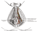
 Pudendal nerve and its course through the pudendal canal (labelled in yellow) | |
| Details | |
| Identifiers | |
| Latin | canalis pudendalis |
| TA98 | A09.5.04.003 |
| TA2 | 2436 |
| FMA | 22071 |
| Anatomical terminology | |
The pudendal canal (also called Alcock's canal) is an anatomical structure formed by the obturator fascia (fascia of the obturator internus muscle) lining the lateral wall of the ischioanal fossa. The internal pudendal artery and veins, and pudendal nerve pass through the pudendal canal, and the perineal nerve arises within it.[1]
Clinical significance
Pudendal nerve entrapment can occur when the pudendal nerve is compressed while it passes through the pudendal canal.[2]
History
The pudendal canal is also known as Alcock's canal, named after Benjamin Alcock.[3]
Additional images
-
 The superficial branches of the internal pudendal artery. (Canal not labeled, but pudendal nerve and internal pudendal artery labeled at bottom right.)

See also
References
![]() This article incorporates text in the public domain from page 421 of the 20th edition of Gray's Anatomy (1918)
This article incorporates text in the public domain from page 421 of the 20th edition of Gray's Anatomy (1918)
- ^ "canalis pudendalis". TheFreeDictionary.com. Retrieved 2023-06-14.
- ^ Chiarioni, Giuseppe; Popa, Stefan-Lucian (2020-01-01), Rao, Satish S. C.; Lee, Yeong Yeh; Ghoshal, Uday C. (eds.), "Chapter 36 - Anorectal pain", Clinical and Basic Neurogastroenterology and Motility, Academic Press, pp. 505–515, doi:10.1016/b978-0-12-813037-7.00036-4, ISBN 978-0-12-813037-7, retrieved 2021-02-08
- ^ Standring, Susan (2016). Gray's anatomy : the anatomical basis of clinical practice (Forty-first ed.). . p. 87. ISBN 9780702052309.
{{cite book}}: CS1 maint: location missing publisher (link)
External links
- Anatomy image: apmalefrontal4-16 at the College of Medicine at SUNY Upstate Medical University
- Cross section image: pelvis/pelvis-e12-15—Plastination Laboratory at the Medical University of Vienna
- Anatomy photo:41:08-0100 at the SUNY Downstate Medical Center — "The Female Perineum: Contents of the Pudendal Canal"
- Diagram at pudendal.info
- Anatomy image:9087 at the SUNY Downstate Medical Center
- Anatomy image:9448 at the SUNY Downstate Medical Center
This anatomy article is a stub. You can help Wikipedia by expanding it. |
Text je dostupný za podmienok Creative Commons Attribution/Share-Alike License 3.0 Unported; prípadne za ďalších podmienok. Podrobnejšie informácie nájdete na stránke Podmienky použitia.
Antropológia
Aplikované vedy
Bibliometria
Dejiny vedy
Encyklopédie
Filozofia vedy
Forenzné vedy
Humanitné vedy
Knižničná veda
Kryogenika
Kryptológia
Kulturológia
Literárna veda
Medzidisciplinárne oblasti
Metódy kvantitatívnej analýzy
Metavedy
Metodika
Text je dostupný za podmienok Creative
Commons Attribution/Share-Alike License 3.0 Unported; prípadne za ďalších
podmienok.
Podrobnejšie informácie nájdete na stránke Podmienky
použitia.
www.astronomia.sk | www.biologia.sk | www.botanika.sk | www.dejiny.sk | www.economy.sk | www.elektrotechnika.sk | www.estetika.sk | www.farmakologia.sk | www.filozofia.sk | Fyzika | www.futurologia.sk | www.genetika.sk | www.chemia.sk | www.lingvistika.sk | www.politologia.sk | www.psychologia.sk | www.sexuologia.sk | www.sociologia.sk | www.veda.sk I www.zoologia.sk